DNA Double Helix Simulation
A free 3D DNA simulation for class: turn the double helix, tap A–T and G–C pairs, build the complementary strand and watch semi-conservative replication.
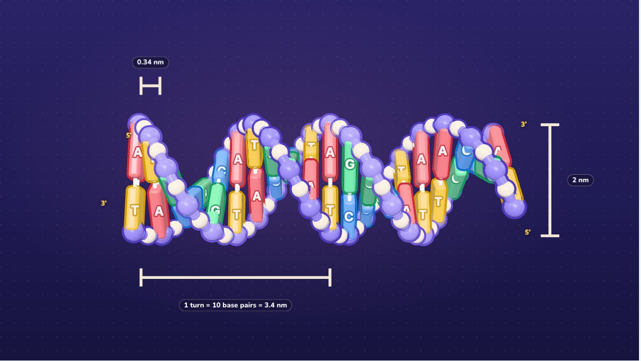
Free, no sign-up, runs in the browser on a projector, TV or touch screen.
Make a review gameFree games library
This is a DNA molecule the whole class can turn in their hands, on one screen. The double helix is built from real measurements: two sugar–phosphate backbones wind round each other, and the bases pair across the middle, adenine with thymine and guanine with cytosine. Tap a pair to see the hydrogen bonds that hold it together, switch on the sizes to see 10 base pairs and 3.4 nm in every turn, and drag the slider to unwind the helix into a flat ladder. Then students build the complementary strand base by base, and watch the molecule copy itself into two, each with one old strand and one new.
How to use it
- Explore: drag to turn the helix, pinch or scroll to zoom. Left alone for a few seconds, it turns slowly by itself. Pick 10, 20 or 30 base pairs, or press New sequence for a different order of bases.
- Tap a base pair: the pair lights up, the rest of the helix dims, and the panel shows the two bases and their hydrogen bonds: two for A–T, three for G–C.
- Sizes: shows one full turn (10 base pairs, 3.4 nm), the distance between two pairs (0.34 nm) and the width (2 nm). Letters shows the base letters and the 5′ and 3′ ends of each strand.
- Ladder ↔ Helix: drag the slider to the left and the helix untwists into a ladder; drag it back and it twists up again.
- Build: the template strand is given along the top. Tap a base in the tray to put it in the glowing gap, or drag it onto any gap. A base that does not fit bounces back with the reason, and the panel counts every base in both strands and checks Chargaff's rule. When every gap is filled, the two strands twist into a helix. New strand gives another template.
- Copy: press Play to watch replication from start to end, or Step to go one base pair at a time while you explain. Old strands are lilac, new strands pink.
- Keyboard and projector: 1, 2 and 3 switch modes. In Explore, the arrow keys turn the helix and pick a pair, and [ and ] unwind and twist. In Build, type A, T, G or C. In Copy, Space plays and pauses and → steps. The full screen button in the top right makes everything bigger for the back row.
What DNA is made of
DNA is a long chain of nucleotides. Each nucleotide has three parts: a sugar (deoxyribose), a phosphate group and one of four bases:
| Base | Letter | Kind | Pairs with | Hydrogen bonds |
|---|---|---|---|---|
| Adenine | A | Purine (two rings, bigger) | Thymine (T) | 2 |
| Thymine | T | Pyrimidine (one ring, smaller) | Adenine (A) | 2 |
| Guanine | G | Purine (two rings, bigger) | Cytosine (C) | 3 |
| Cytosine | C | Pyrimidine (one ring, smaller) | Guanine (G) | 3 |
The sugars and phosphates link up into a backbone on the outside of each strand (the lilac tubes with white beads in the model). The bases point inwards, and each one pairs with the base opposite it on the other strand.
Base pairing and Chargaff's rule
A always pairs with T, and G always pairs with C. Each pair is one big base (a purine) and one small base (a pyrimidine), so every rung of the ladder is the same width. Two purines would be too wide, two pyrimidines too narrow, and A with C or G with T is the right size but cannot form the hydrogen bonds. Build mode explains exactly this when a base bounces back.
Because every A has a T opposite it and every G has a C, a double strand always has as many A as T and as many G as C: this is Chargaff's rule. A + G (purines) = T + C (pyrimidines) = half the bases. If a DNA sample is 30% A, it is also 30% T, and the other 40% is split into 20% G and 20% C.
The hydrogen bonds holding the two strands together are counted the same way:
H = 2 × (number of A–T pairs) + 3 × (number of G–C pairs)
The template ATGCGTACCA in Build has 5 A–T and 5 G–C pairs, so 2 × 5 + 3 × 5 = 25 hydrogen bonds. DNA rich in G and C holds together more tightly.
The double helix in numbers
The model uses the measurements of B-DNA, the usual form in cells:
- about 10 base pairs in every full turn, each pair turned 36° from the one before;
- 0.34 nm between neighbouring pairs, so one turn is 3.4 nm long;
- 2 nm across.
So a stretch of DNA with N base pairs makes N ÷ 10 turns and is N × 0.34 nm long. The 20 pairs in Explore make 2 turns and 6.8 nm. Human chromosome 1 has about 248 million base pairs: 248,000,000 × 0.34 nm is about 8.4 cm of DNA, folded into a nucleus a few thousandths of a millimetre across.
The two strands run in opposite directions (they are antiparallel): one reads 5′ to 3′ from left to right, the other 5′ to 3′ from right to left. That is why the complementary strand of 5′-ATGC-3′ is written 3′-TACG-5′, or 5′-GCAT-3′ when read from its own 5′ end.
Replication: one molecule becomes two
Before a cell divides, it copies its DNA. Copy mode shows the steps:
- The double helix unwinds.
- Helicase unzips the two strands by breaking the hydrogen bonds between the bases.
- Free nucleotides pair with the bases of each old strand, A with T and G with C, so each old strand is the template for a new one.
- DNA polymerase joins the new nucleotides into a strand.
- The two new molecules twist up. Each has one old strand and one new strand, and both are identical to the first molecule.
Keeping one old strand in each copy is called semi-conservative replication. Meselson and Stahl showed it in 1958 by growing bacteria on heavy nitrogen and then on light nitrogen: after one round every DNA molecule was half heavy, half light.
Try this
- Tap any A–T pair, then any G–C pair. Count the hydrogen bonds. Which pair is harder to pull apart?
- Switch to 10 base pairs with Sizes on. How many turns? Now 30: predict the turns and the length before you look at the panel.
- In Build, put G opposite a template A, then C opposite a template T. Read why each one bounces back.
- Finish a strand and check the counts. Is A = T in the template strand alone? In both strands together?
- In Copy, stop halfway. Which strands are old and which are new? Predict how many old strands there will be after a second round of copying.
Teaching ideas
The simulation fits the DNA units of high school biology: the structure of DNA, complementary base pairing and replication, as in the AP Biology unit on gene expression and regulation, GCSE Biology's genome topic and IB Biology's DNA structure and replication. In the NGSS it supports HS-LS1-1 (DNA and the structure of proteins) and HS-LS3-1 (DNA and chromosomes in inheritance).
- Warm-up (3 minutes): show the helix turning, with letters off. Ask: "What do you think holds the two strands together?"
- Base pairs (7 minutes): students come to the board and tap pairs. Ask: "Why does A never pair with G?" Use the Build hints to talk about size and hydrogen bonds.
- Numbers (5 minutes): turn on Sizes and work out turns and length for 10, 20 and 30 base pairs, then for a whole chromosome.
- Build in teams (10 minutes): each team builds a complementary strand at the board while the others check with the counts and Chargaff's rule.
- Replication (8 minutes): step through Copy, one pair at a time. Ask the class to predict each new base before you press Step.
- Review: make a review game: the whole class plays on the same screen in teams, with your phone as the remote.
Questions teachers ask
Why does A pair with T, and G with C?
Each pair has one big base with two rings (A or G) and one small base with one ring (T or C), so every rung is the same width and the helix stays smooth. Within that, only A and T line up to make two hydrogen bonds, and only G and C to make three. A with C is the right width but its atoms don't line up to bond.
Is a turn really exactly 10 base pairs?
Close. Textbooks use the round numbers from the first X-ray pictures: 10 base pairs and 3.4 nm a turn. Measured in water, B-DNA has about 10.5 base pairs a turn. The simulation uses 10, the number students will see in exams.
What do 5′ and 3′ mean?
They name the two ends of a strand, after the carbon atoms of the sugar (the 5th and the 3rd). A strand has a direction, like an arrow, and the two strands of a helix point opposite ways. New DNA is always built from the 5′ end toward the 3′ end.
Does replication in a real cell look like this?
The main steps are the same, but the model leaves out details. In a cell, a short RNA primer starts each new strand, and because both new strands can only grow from 5′ to 3′, one of them (the lagging strand) is built in short pieces called Okazaki fragments that are then joined. Copying also starts at many points along a chromosome at once. The simulation draws both new strands growing smoothly so the main idea stands out: two copies, each with one old strand.
Is the model to scale?
In Explore, yes: the helix uses the real width, rise and turn. In Build and Copy the flat ladder is stretched so every base is big enough to tap and read on a board, and the helices they twist into are a little stretched too.
Can students use it on an interactive whiteboard?
Yes. Every finger drags its own base in Build, so two students can work at once, and the bases in the tray are big enough to tap. On a phone the helix stands upright and the panel sits underneath. Nothing to install and no sign-up: it runs in the browser and it's free.